It became apparent that epigenetic pathways play an important role in the reproduction of plants, for example in the regulation of flowering time and in the development of the female gametophyte. The timing of flowering is crucial for the reproductive success of the plants, as it ensures that flowering occurs at the best time for pollination and seed set. Likewise, cell fate decisions during female gametophyte development are important for the formation of gametes, and the chromatin states of the gametes affect seed development after fertilization.
Epigenetic pathways are mechanisms that regulate gene expression without altering the underlying DNA sequence. These pathways involve modifications to DNA and histone proteins, as well as the action of regulatory small non-coding RNAs (sncRNAs) and long non-coding RNAs. Regulatory sncRNAs, including miRNAs and siRNAs, control gene expression by interacting with proteins, primarily members of the Argonaute (AGO) protein family. They guide the AGOs to specific DNA or RNA targets for transcriptional or post-transcriptional gene silencing. While transcriptional gene silencing is achieved through epigenetic modifications like DNA methylation or histone modifications, post-transcriptional gene silencing involves the destruction or suppression of translation of the target mRNA(s).
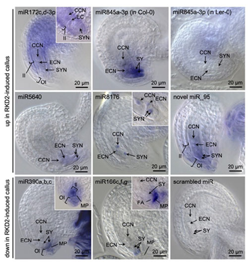
Our research in this project aims to investigate the role of novel small non-coding RNAs that we have previously identified (Sprunck et al., 2019) and their putative target genes. In addition, we are studying those AGOs expressed in Arabidopsis egg cells (Sprunck et al., 2019) and previously identified AGO interactors to elucidate their role in plant reproduction.